Blab Tech
IIT Madras Releases First Cell-Resolution 3-D Atlas of the Human Brainstem
Scientists at the Indian Institute of Technology Madras (IIT‑Madras) have unveiled ANCHOR, the world’s most detailed three‑dimensional map of the human brainstem at cellular resolution. Announced on Friday, the atlas charts more than 200 distinct structures—from nuclei to fibre tracts—throughout the brainstem’s lifespan, from prenatal development to adulthood.
The brainstem, a slender stalk that links the cerebrum to the spinal cord, governs essential functions such as breathing, sleep, wakefulness and basic motor activity. When it is damaged, severe neurological disorders often follow. Yet clinicians have long worked with broad, low‑resolution maps that cannot identify which specific cells are affected.
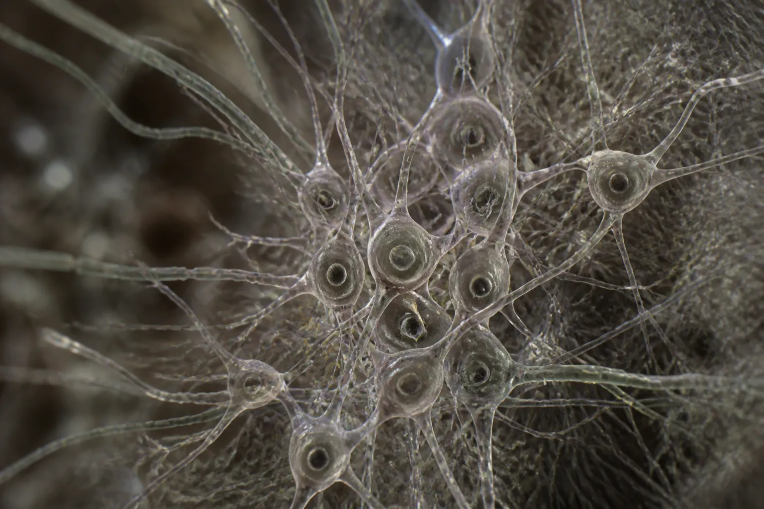
ANCHOR overcomes this limitation by fusing high‑field magnetic resonance imaging (MRI) with detailed histological and neurochemical data. Researchers applied eight different immunostains—antibody‑based dyes that bind to specific proteins—to several hundred thin tissue slices. The staining highlights individual cell types, allowing the atlas to resolve structures at the level of single cells.
The atlas functions like a “Google Earth” for the brainstem. A clinician can start with a conventional MRI scan, then zoom progressively through tissue layers, fibre tracts, and finally down to individual cells. This multi‑scale approach delivers a continuous, navigable view of the brainstem’s anatomy and chemistry.
According to the Sudha Gopalakrishnan Brain Centre at IIT‑Madras, ANCHOR is the first atlas to integrate MRI, histology and neurochemical mapping in a single, freely accessible resource. The atlas is available at anchor.humanbrain.in and can be accessed by researchers and clinicians worldwide at no cost.
Ajay Kumar Sood, principal scientific adviser to the Government of India, said that the maps will help identify specific cell populations affected in brainstem lesions, a development he described as “critical for clinical applications.” The ability to see which cells are damaged could improve diagnosis, guide surgical planning, and inform targeted therapies for conditions such as brainstem stroke, neurodegenerative disease and congenital malformations.
The project’s scope is unprecedented. It includes over 200 brainstem nuclei and fibre tracts, covering the midbrain, pons and medulla oblongata. The atlas spans the full human lifespan, providing developmental context that is currently missing from most brainstem studies.
IIT‑Madras’s announcement follows a series of publications that describe the atlas’s construction and validation. The team used a combination of high‑resolution MRI scans and serial immunostaining to build a comprehensive, 3‑D reconstruction of the brainstem’s cellular architecture.
The release of ANCHOR is expected to accelerate research into brainstem disorders. By offering a precise, cell‑level reference, the atlas can serve as a baseline for comparative studies, help validate computational models, and support the development of new diagnostic tools.
While the atlas is a significant advance, its creators note that further work is needed to extend the resource to diseased brains and to integrate functional data. Nonetheless, the availability of a free, high‑resolution brainstem atlas represents a milestone for neuroscience and clinical neurology.
The Sudha Gopalakrishnan Brain Centre plans to expand ANCHOR by adding additional developmental stages and incorporating data from patients with brainstem pathology. The institute also intends to collaborate with international partners to refine the atlas’s annotations and to develop user tools that facilitate its integration into clinical workflows.
In summary, ANCHOR provides the first publicly available, cell‑resolution 3‑D atlas of the human brainstem, combining MRI, histology and immunostaining to map over 200 structures across the lifespan. The resource is poised to improve the precision of neurological diagnosis and to support research into brainstem function and disease.
The brainstem, a slender stalk that links the cerebrum to the spinal cord, governs essential functions such as breathing, sleep, wakefulness and basic motor activity. When it is damaged, severe neurological disorders often follow. Yet clinicians have long worked with broad, low‑resolution maps that cannot identify which specific cells are affected.
ANCHOR overcomes this limitation by fusing high‑field magnetic resonance imaging (MRI) with detailed histological and neurochemical data. Researchers applied eight different immunostains—antibody‑based dyes that bind to specific proteins—to several hundred thin tissue slices. The staining highlights individual cell types, allowing the atlas to resolve structures at the level of single cells.
The atlas functions like a “Google Earth” for the brainstem. A clinician can start with a conventional MRI scan, then zoom progressively through tissue layers, fibre tracts, and finally down to individual cells. This multi‑scale approach delivers a continuous, navigable view of the brainstem’s anatomy and chemistry.
According to the Sudha Gopalakrishnan Brain Centre at IIT‑Madras, ANCHOR is the first atlas to integrate MRI, histology and neurochemical mapping in a single, freely accessible resource. The atlas is available at anchor.humanbrain.in and can be accessed by researchers and clinicians worldwide at no cost.
Ajay Kumar Sood, principal scientific adviser to the Government of India, said that the maps will help identify specific cell populations affected in brainstem lesions, a development he described as “critical for clinical applications.” The ability to see which cells are damaged could improve diagnosis, guide surgical planning, and inform targeted therapies for conditions such as brainstem stroke, neurodegenerative disease and congenital malformations.
The project’s scope is unprecedented. It includes over 200 brainstem nuclei and fibre tracts, covering the midbrain, pons and medulla oblongata. The atlas spans the full human lifespan, providing developmental context that is currently missing from most brainstem studies.
IIT‑Madras’s announcement follows a series of publications that describe the atlas’s construction and validation. The team used a combination of high‑resolution MRI scans and serial immunostaining to build a comprehensive, 3‑D reconstruction of the brainstem’s cellular architecture.
The release of ANCHOR is expected to accelerate research into brainstem disorders. By offering a precise, cell‑level reference, the atlas can serve as a baseline for comparative studies, help validate computational models, and support the development of new diagnostic tools.
While the atlas is a significant advance, its creators note that further work is needed to extend the resource to diseased brains and to integrate functional data. Nonetheless, the availability of a free, high‑resolution brainstem atlas represents a milestone for neuroscience and clinical neurology.
The Sudha Gopalakrishnan Brain Centre plans to expand ANCHOR by adding additional developmental stages and incorporating data from patients with brainstem pathology. The institute also intends to collaborate with international partners to refine the atlas’s annotations and to develop user tools that facilitate its integration into clinical workflows.
In summary, ANCHOR provides the first publicly available, cell‑resolution 3‑D atlas of the human brainstem, combining MRI, histology and immunostaining to map over 200 structures across the lifespan. The resource is poised to improve the precision of neurological diagnosis and to support research into brainstem function and disease.